
Capnography During Intrahospital Transport
Myron Yaster MD, Lynn Martin MD MBA, Conor Mc Donnell MD MB

Back when I was in college, my friends and I would argue endlessly over the question: “If you were stranded on a desert island and could have only one (vinyl) album (OK for most of you, that’s now a streamed electronic album) what would it be”? Beatles or the Stones? Hendrix or Garcia? Davis or Bird? Ella or Billie? You get the idea, so what’s the desert island anesthesia monitor? Pulse oximeter or Capnograph”? For many (most?) of you, I think it’s pulse oximetry. For me it’s capnography. (see an old PAAD here )
From a previous reader response: “There’s no question that pulse oximetry is a valuable monitoring tool in any clinical situation associated with hypoxemia. As such, changes in SpO2 are a sensitive indicator of disease severity and response to treatment in conditions associated with ventilation/perfusion mismatch and intrapulmonary or intracardiac shunt, such as asthma, bronchopulmonary dysplasia, bronchiolitis, pneumonia, and congenital heart disease. However, conditions associated with hypercarbia due to reduced alveolar ventilation (hypoventilation) as a result of partial airway obstruction or depression of ventilation (endotracheal tube kinking or disconnect, ventilator failure, drugs) will not be associated with significant changes in SpO2, particularly if supplemental oxygen is administered, until partial pressure of carbon dioxide in arterial blood is extremely elevated. Hence, use of pulse oximetry could result in a delay of diagnosis in these conditions – especially in the PACU or PICU or in sedated patients in whom oxygen supplementation is standard practice. Moreover, there is only one monitor that we use than can singlehandedly provide data about all 3 of the ABCs: Capnography. If any one of airway, breathing, or circulation are not intact, there is a change (or loss) of EtCO2.” (Justin L. Lockman MD MSEd)
In today’s PAAD, researchers at the Boston Children’s Hospital(1) using a quality improvement (QI) initiative, instituted the mandatory use of capnography in intrahospital transport of mechanically ventilated pediatric patients with congenital heart disease (CHD). They describe how their multidisciplinary QI team, consisting of pediatric cardiac anesthesiologists and anesthesia technicians, implemented the initiative using multiple Plan–Do–Study–Act (PDSA) cycles in accordance with the Standards for Quality Improvement Reporting Excellence (SQUIRE) guidelines. The senior authors, Drs. Jim DiNardo and Viviane Nasr, are members of the PAAD’s executive council and are frequent contributors to the PAAD. Myron Yaster MD
Original article
Brown ML, Whiting D, DiNardo JA, Nasr VG. Institutional Experience Introducing Portable Capnography Intrahospital Transport of Ventilated Patients With Congenital Heart Disease. Anesth Analg. 2026 Aug 1;143(2):421-423. doi: 10.1213/ANE.0000000000007940. Epub 2026 Jan 14. PMID: 41534096.
Intrahospital transport of mechanically ventilated pediatric patients with congenital heart disease (CHD) is a high-risk process associated with significant respiratory and hemodynamic instability.(2) (see previous PAAD here ) In this QI study, Brown et al. describe the successful introduction of portable capnography for intrahospital transport of ventilated pediatric cardiac patients at a high-volume tertiary referral center, demonstrating that systematic implementation can achieve improvements in monitoring compliance. The authors failed to state their SMART (Specific, Measurable, Attainable, Relevant, and Time-bound) aim or target for the improvement efforts. In Seattle we typically strive for >80% to achieve a reliable process. We call the improvements sustained when the improvements have been maintained for at least 12 consecutive time intervals (i.e., weeks, months). Do we stop there? What is standing in the way for high reliability? It is people, equipment availability, etc.? We (LDM and CM) pursue continuous improvements using additional Plan–Do–Study–Act (PDSA) cycles to reach >90%, then >95%, and even >98% compliance to achieve highly reliable systems state. At these levels, it truly becomes the culture, ‘the way things are done around here’.
The authors identified the absence of end-tidal CO₂ (EtCO₂) monitoring during transport as an institutional practice gap. As you all know and as discussed in Myron’s introduction, capnography provides continuous, noninvasive assessment of ventilation, pulmonary perfusion, and metabolism while confirming endotracheal tube position and detecting ventilation-related complications. A multidisciplinary QI team consisting of pediatric cardiac anesthesiologists and anesthesia technicians implemented the initiative using multiple PDSA cycles in accordance with the Standards for Quality Improvement Reporting Excellence (SQUIRE) guidelines. Educational sessions, equipment evaluation, workflow redesign, electronic medical record documentation, and iterative process refinement were incorporated throughout implementation. Figure 1 from the article illustrates the progressive introduction of the technology, including equipment selection, staff education, workflow integration, relocation of monitors, and subsequent equipment modifications to improve availability and reduce device loss.
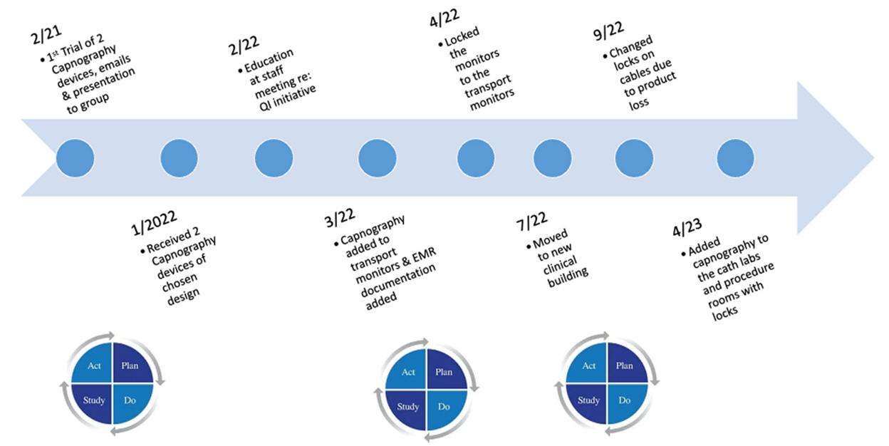
Brown et al. reviewed all intubated CHD patients transported by the anesthesia service between January 2022 and May 2024. During the study period, 2,996 mechanically ventilated patients underwent intrahospital transport, with complete monitoring data available for 96% of transports. The median patient age was one year, and nearly half of the cohort consisted of neonates younger than 30 days, emphasizing the high-risk nature of the population. Approximately three-quarters of transports originated in the operating room, with the remainder occurring after cardiac catheterization or advanced cardiac imaging.
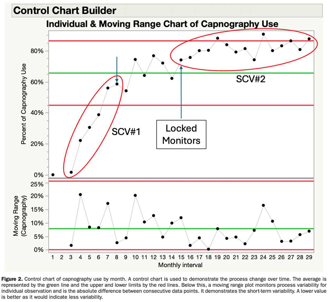
The initiative resulted in improvement >80% in capnography uses for the last 6 months. During the final three months of observation, monitoring compliance reached 86.3%, 80.7%, and 87.6%, demonstrating reliable adoption across the anesthesia service. Figure 2 from the article is an XmR (Mean and moving Range) Control chart which illustrates progressive improvement in compliance over successive implementation cycles.
The use of Statical Process Control (SPC) methods and control charts with their control limits is a significant step forward in understanding process change over the more typically used run chart. (3) Control charts allow users to distinguish between common cause (random) and special cause variation (SCV). There are well established rules use to define SCV as part of SPC methods. In quality improvement initiatives, the team is looking for SCV on the charts. Below I (LDM) have modified their original figure 2 to indicate at target of 80% compliance and identified two SCV signals on their chart. The first SCV (six consecutive increasing points) occurs with the introduction of the capnometers. The second SCV (eight or more consecutive point above the mean) occurred after the locking of monitors. Using SPC methods, this chart should have three separate cohorts (broken at the arrows) each with unique means and control limits. Note: There appears to be no SCV signal in the moving range (variability) chart if the highlighted point is on the mean as it appears.
Using the departmental event self-reporting system, only two transport-related adverse events or near misses occurred during the study period, neither of which was attributable to the capnography device itself. Instead, both events were associated with physiologic changes occurring during transition from mechanical ventilation to manual ventilation. Although the study was not powered to demonstrate improved clinical outcomes, these findings suggest that portable capnography can be integrated safely into routine transport without introducing additional risk. One can speculate if more adverse events would have been identified with ‘button tags’ (i.e., code, CPR, etc.), ‘trigger medication’ (i.e., epinephrine, etc.), or other sources of information (i.e., handoff reports) for additional adverse events, enhancing the ‘believability’ of this balance measure.
Implementation was associated with additional financial considerations. The acquisition cost was approximately US$2,087 per monitor, with disposable adapters costing US$10–16 per patient, resulting in estimated disposable expenditures of US$24,000–47,000 during the study period. However, reuse of disposable adapters by the intensive care unit frequently reduced overall institutional costs. The authors identified equipment availability and cost as the principal barriers to universal implementation.
This is a very reasonable fix to a common problem. However, we wonder why universal capnography monitoring should be limited to the intrahospital transport of only INTUBATED CHD patients? Why not use capnography in ALL intubated patients? This could be the next phase of their continuous improvement efforts enhancing safety. The final and most difficult task would be to use capnography in all intubated and non-intubated patients during transpotrt (think PACU)? Airway obstruction is common during transport of many of our patients. You’ve all experienced this in transport of non-intubated, semi-or unconscious patients to the PACU. Indeed, Dr. Peter Safar, widely recognized as the father of CPR, revolutionized emergency medicine by discovering that in unconscious humans, flexing the neck caused airway obstruction due to relaxed soft tissues and the tongue naturally falling backward. His landmark research in the 1950-60s established the foundational ABC’s of resuscitation. Technical limits in non-intubated patients and costs escalate with this last step and could represent ‘a bridge too far’.
How do you monitor intubated and non-intubated patients during intrahospital transport? Do you routinely use pulse oximetry? Capnography? ECG? Send your thoughts and comments to Myron (myasterster@gmail.com) and he will post in a Friday reader response
References
1. Brown ML, Whiting D, DiNardo JA, Nasr VG. Institutional Experience Introducing Portable Capnography Intrahospital Transport of Ventilated Patients With Congenital Heart Disease. Anesthesia and analgesia. 2026;143(2):421–3. Epub 20260114. doi: 10.1213/ane.0000000000007940. PubMed PMID: 41534096.
2. Haydar B. Error Traps in the Intrahospital Transport of Critically Ill and Anesthetized Children. Paediatric anaesthesia. 2025;35(7):497–503. Epub 20250408. doi: 10.1111/pan.15112. PubMed PMID: 40198097; PubMed Central PMCID: PMC12149488.
3. Provost LP, Murray SK. The Health Care Data Guide: Learning from Data for Improvement (John Wiley and Sons; Hoboken, NJ, 2022).